A crystal structure, ten docking poses, and an explicit validation gap
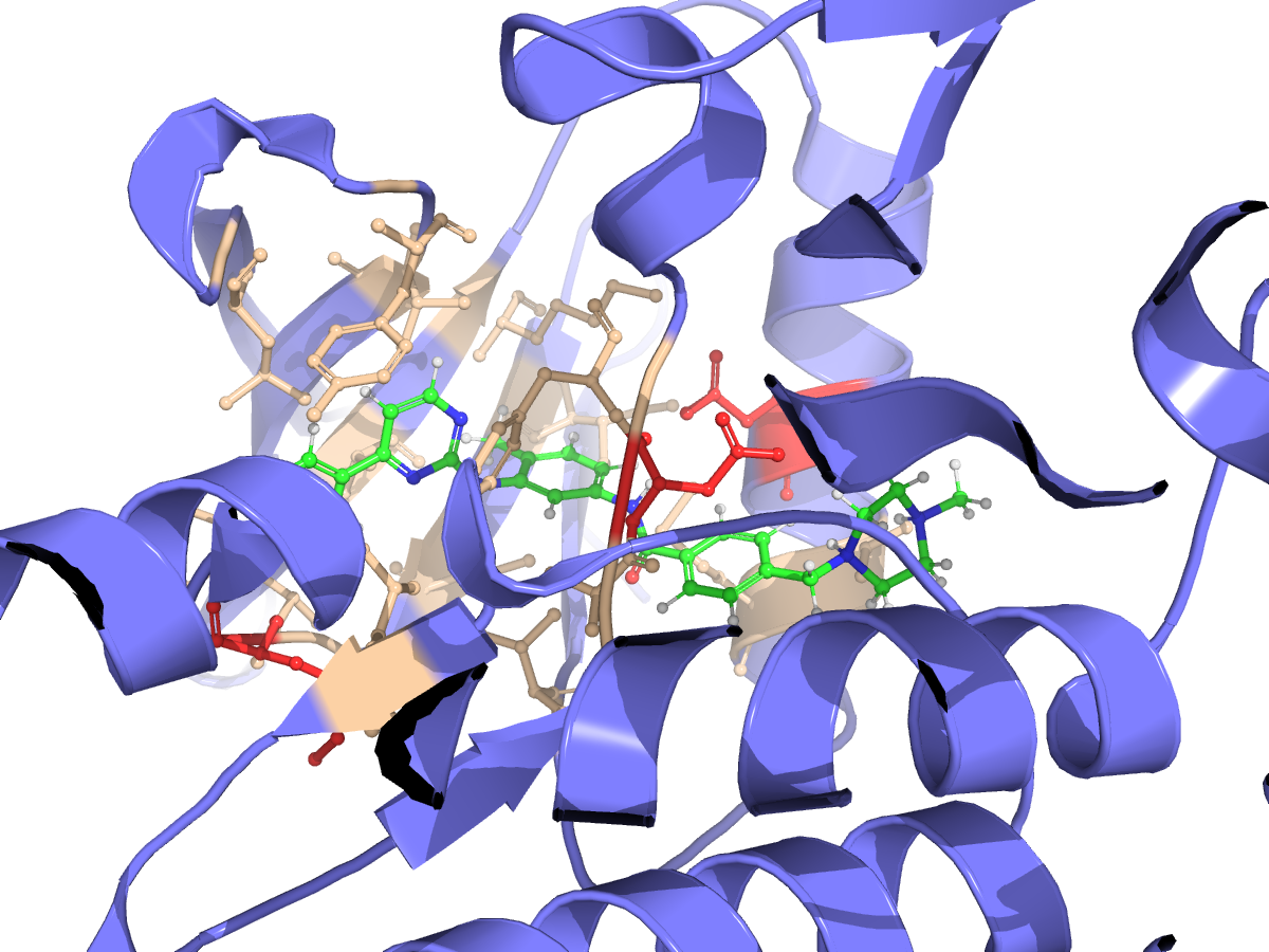
Session 3b376bba used the ABL1 kinase crystal structure 2HYY, chain A, with crystallographic imatinib retained as a site reference before receptor cleaning. Vina generated ten poses; ProLIF characterized their contacts. Docking completed, but the heavy-atom RMSD comparison to the crystal ligand did not. These results describe the reported run, not a validated recovery of the experimental pose.
Can the workflow dock imatinib into a known ABL1 binding site?
The session requested structure retrieval, receptor cleaning, pocket detection, ligand preparation, docking, and a visualization of the resulting pose. The source was the 2HYY crystal structure, reported at 2.4 Å resolution, with chain A selected from four kinase-domain copies.
What poses, Vina scores, and residue contacts does imatinib produce in the cleaned 2HYY chain A receptor, and can the workflow assess pose recovery against crystallographic imatinib?
| Session | 3b376bba; analysis dated September 6, 2026 |
|---|---|
| Receptor source | RCSB PDB 2HYY; chain A |
| Ligand input | User-supplied imatinib SMILES; 37 heavy atoms reported |
| Site reference | Crystallographic imatinib STI:A:600 |
| Pocket detection | fpocket: 17 sites; pocket 1 selected |
| Docking and contacts | AutoDock Vina 1.1.2; ProLIF 2.2.1 / MDAnalysis 2.10.0 |
From structure retrieval to pose inspection
| Box center (Å) | 14.878, 16.836, 17.241 |
|---|---|
| Box dimensions (Å) | 25.9 × 23.6 × 18.8 |
| Box source | STI:A:600 coordinates plus 5 Å padding |
| Site coverage | 37/37 reference ligand atoms; 491 receptor atoms enclosed |
| fpocket pocket 1 | Score 0.894; reported volume 474 ų; 5.02 Šfrom STI centroid |
Ten poses, with a top Vina score of −11.9 kcal/mol
Ten returned poses scored from −11.9 to −9.0 kcal/mol in this receptor and search box.
ProLIF recorded 691 interaction occurrences across ten poses, including 67 for the top pose.
ASP381, MET290, and PHE382 had reported hydrophobic contacts in all ten poses.
The reference-ligand match failed; no heavy-atom redocking RMSD was obtained.
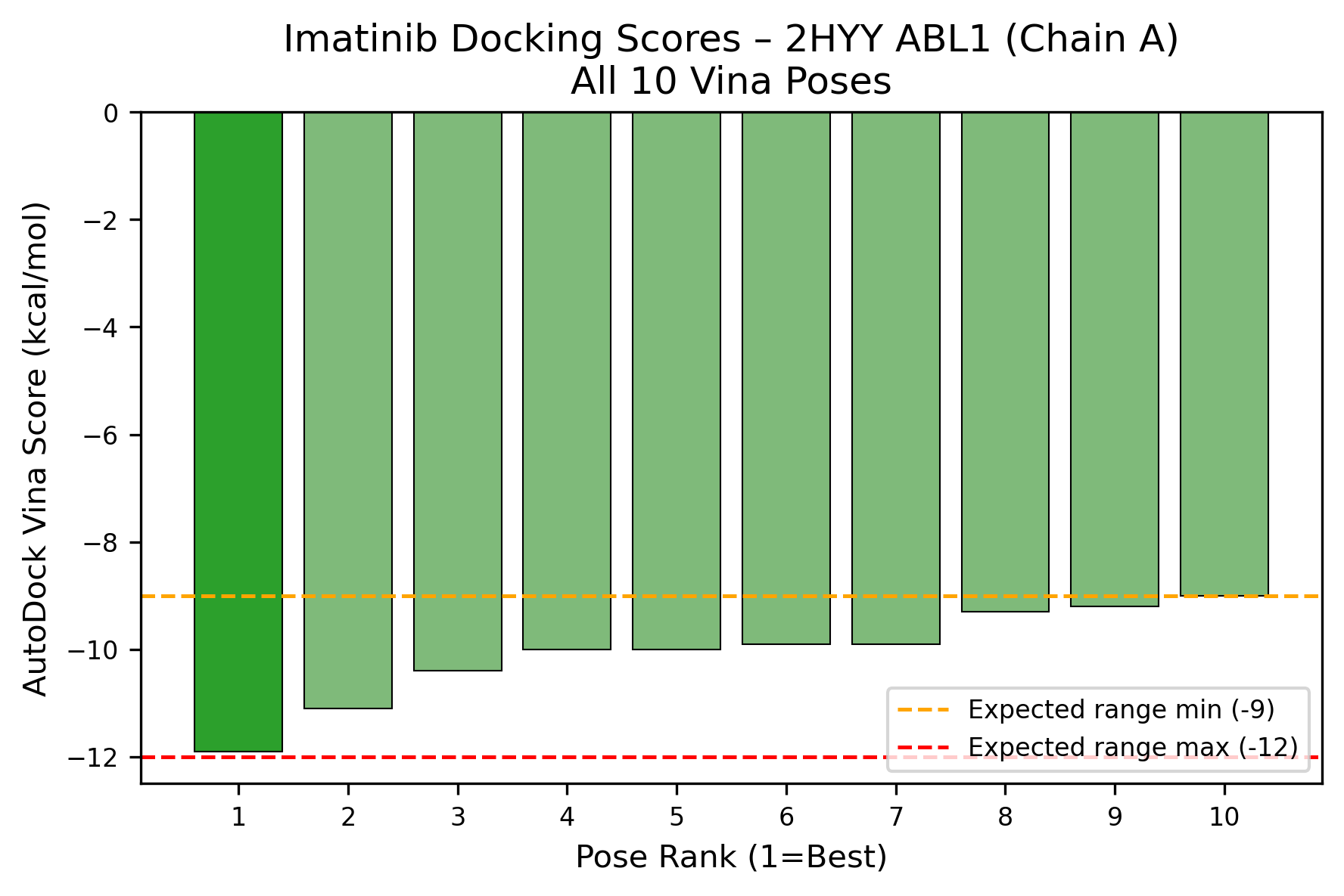
| Pose rank | Vina score (kcal/mol) |
|---|---|
| 1 | −11.9 |
| 2 | −11.1 |
| 3 | −10.4 |
| 4 | −10.0 |
| 5 | −10.0 |
| 6 | −9.9 |
| 7 | −9.9 |
| 8 | −9.3 |
| 9 | −9.2 |
| 10 | −9.0 |
Which contacts recur across the docked poses?
ProLIF contact frequencies summarize how many of the ten generated poses contain each residue–interaction pair. They describe this docking ensemble; they are not time occupancies from molecular dynamics or probabilities of binding.
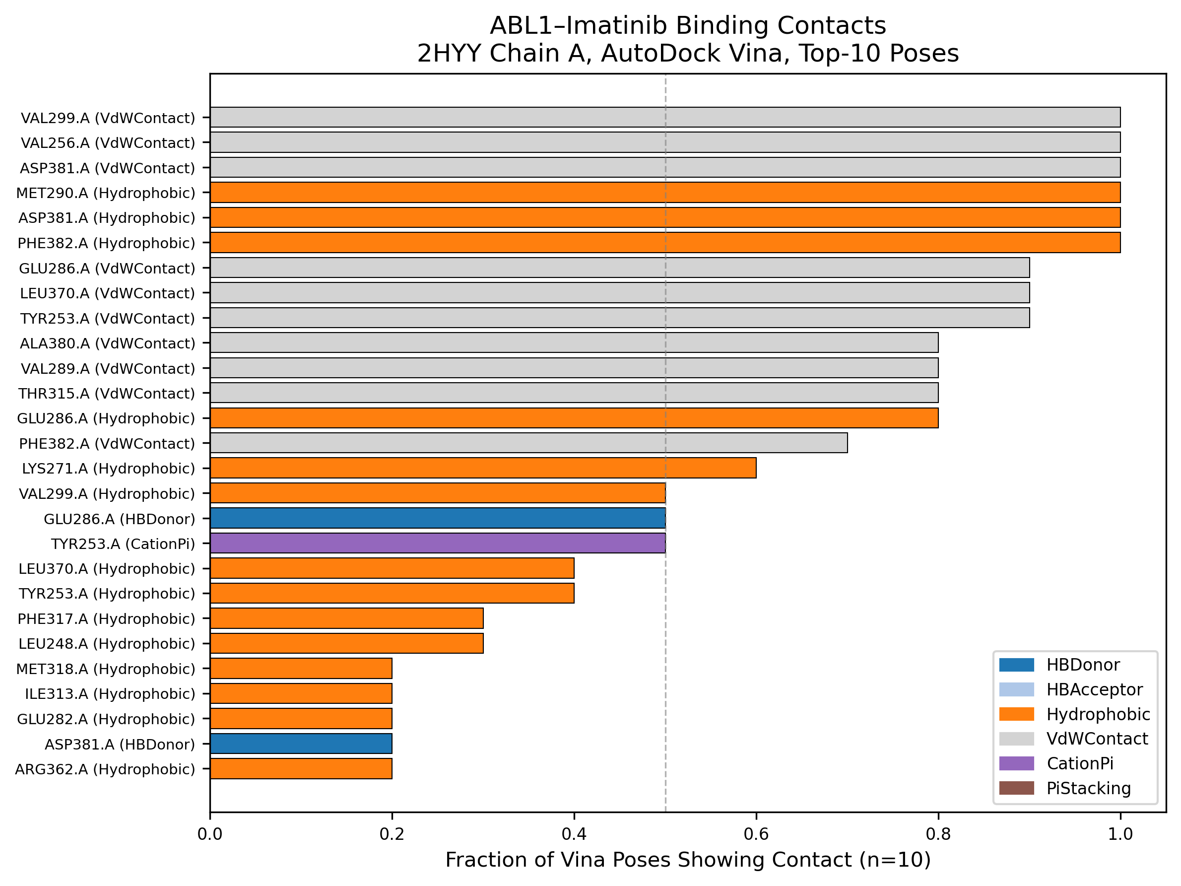
| Residue (chain A) | Reported interaction | Poses with contact |
|---|---|---|
| ASP381 | Hydrophobic | 10/10 |
| MET290 | Hydrophobic | 10/10 |
| PHE382 | Hydrophobic | 10/10 |
| GLU286 | Hydrophobic | 8/10 |
| THR315 | VdWContact | 8/10 |
| GLU286 | HBDonor | 5/10 |
| TYR253 | CationPi | 5/10 |
The docking run completed; pose recovery remains unvalidated
- The heavy-atom RMSD calculation failed with “No sub-structure match found.” The report attributes this to a mismatch between the prepared protonated ligand and the crystal reference; the actual pose error remains unknown.
- The report flags the protocol as docking_protocol_unvalidated. A favorable score or a visually plausible pose does not replace a successful comparison with the experimental ligand coordinates.
- The receptor is rigid, with crystal waters removed. Side-chain relaxation, backbone movement, and water-mediated contacts were not assessed.
- Protonation is uncertain. The report describes a doubly protonated piperazine and an Open Babel warning about simultaneous -p/-h flags, so the preparation should not be treated as a verified pH-specific state.
- One receptor chain and one random seed were used. Sensitivity to other chains, seeds, or preparation choices was not assessed.
- Vina scores are exploratory ranking values. This run does not establish binding affinity, potency, selectivity, or resistance effects.
A next validation step is to reconcile ligand identity, protonation, and atom mapping before repeating the crystal-pose RMSD comparison. Preparation and seed sensitivity can then be examined with the same receptor and scoring protocol.
The session report and its recorded outputs
The downloadable PDF preserves session 3b376bba, including the request, plan, methods, scores, figures, limitations, and an exported reasoning excerpt. It was generated on September 10, 2026. The files below are listed in that report; this page provides the PDF and extracted figures, not a separately downloadable raw analysis bundle.
2hyy.cif / 2HYY_chainA.pdbRetrieved structure and selected receptor chainimatinib.csvLigand SMILES inputfpocket_analysis.jsonPocket rankings and reference-site distancesdocking_scores.csv / docking_box.jsonPose scores and executed search-box geometrydocking_summary.json / qc_metrics.jsonDocking settings, summary, and quality flagsredock_validation.jsonFailed RMSD validation attemptinteraction_details.csv / residue_frequency.csvPer-pose contacts and contact frequenciespose_0000.pdbqt / pose_0000.sdfDocked pose files listed in the report2HYY_imatinib_docked.psePyMOL session listed with the report figures